Velvet fish disease can occur in both marine and freshwater systems. Although the marine form is much more common, fish owners should be aware of the clinical signs associated with this disease and what to do if you suspect velvet fish disease is in your system.
Clinical Signs of Velvet in Fish
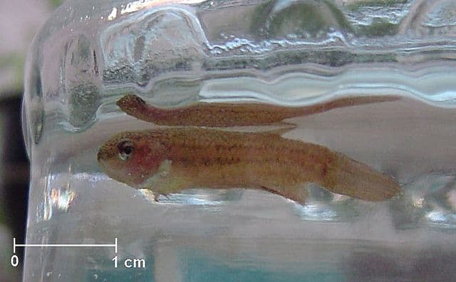
For both freshwater and marine, the clinical signs of fish velvet disease are identical, even though the causative parasite is distinctly different. Infected fish will appear to have gold to brown “dust” sprinkled all over their external body. They will likely show secondary signs of skin irritation, through bruising, missing scales, redness on the body or “flashing” behavior.
With a life cycle almost identical to White Spot Disease (I. multifiliis), infective stages can be seen on both the skin and gills. With the destruction of normal gill tissue, fish will become lethargic, have increased respirations or respiratory effort, and may not be able to maintain normal body position. Mildly infected individuals will stay in areas of higher oxygenation, such as by filter returns, waterfalls, powerheads and other outflows.
Additional clinical signs include decrease or no appetite, hiding or isolating, or sudden death. As with many other parasitic infections, secondary bacterial and fungal infections may be concurrent.
Freshwater Velvet Fish Disease
In freshwater, velvet fish disease is caused by the protozoan parasite, Piscinoodinium. This disease is more common in warmer water, with anabantids (ex. gouramis and bettas), cyprinids (ex. barbs) and cyprinodontids (ex. killifish) being more commonly affected. It can also affect aquaculture species, such as tilapia, and is associated with high mortalities secondary to sudden temperature drops.
Marine Velvet Fish Disease
The marine variant of velvet disease in fish is caused by Amyloodinium. It’s life cycle and presentation is identical to its freshwater variant despite not being a close genetic relative of Piscinoodinium. This disease is significantly more common in marine systems than freshwater systems. Just one infective tomont can produce 64-256 infective dinospores. The dinospores develop into trophonts which is the “dust” that you can see with the naked eye on your fish. The rate of multiplication is optimized between 73 to 81F (23 to 27C). This means that a system can become easily overwhelmed very quickly if the parasite is not caught quickly and effectively treated.

How to Treat Velvet Disease
Since it can appear very similar to white spot disease, a correct diagnosis is critical to successful treatment. Due to the difference in reproductive cycles between the two infectious protozoans, treatment must be long enough to cover all life cycles. If you suspect your fish is sick, contact your local aquatic veterinarian ASAP. Do not run the risk of an incorrect diagnosis and treatment! Delay in correctly identifying the infective pathogen can result in a tank or pond full of dead fish. We understand that using a qualified veterinarian is more expensive than grabbing a box of unknown medication off the shelf, but you are guaranteed to be more likely to save your fish.
How to Prevent Velvet Disease in Fish
As with all infectious diseases, proper quarantine of all new additions is critical to preventing the spread of fish velvet disease. No matter where you purchase your fish from or what the vendor promises, no one can guarantee a “disease free” fish addition. Fish are living animals and cannot be sterilized. Set up a proper quarantine tank prior to the new additions arrival and stick to a conservative quarantine protocol. Remember, all pathogens are temperature dependent! Protocols will vary with your fish species and their optimum temperature. Do not try to speed up quarantine by increasing your water temperature! This may change how different pathogens replicate and give you a false sense of health.
